Ultrasonography (USG)
The FORTEMED Medical Centre offers a wide range of ultrasound examinations:
General ultrasonography
Ultrasound is a quick, painless, informative, and harmless diagnostic imaging technique used to visualise organs and tissues. Ultrasonography at the Fortemed Medical Centre is performed using state-of-the-art equipment with the latest-generation sensors and software, which deliver higher image resolution and, therefore, greater accuracy.
The FORTEMED ultrasound diagnostics team
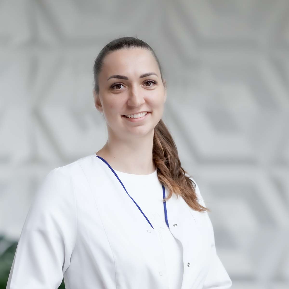 Monta Urbāne
gastroenterologist, ultrasonography of the abdominal cavity
A gastroenterologist who provides patients with a comprehensive approach to health care - from detailed consultation to accurate diagnosis. Abdominal ultrasonography is performed, the symptoms and visual picture are carefully evaluated, helping to establish an accurate diagnosis and choose the most effective treatment.
Monta Urbāne
gastroenterologist, ultrasonography of the abdominal cavity
A gastroenterologist who provides patients with a comprehensive approach to health care - from detailed consultation to accurate diagnosis. Abdominal ultrasonography is performed, the symptoms and visual picture are carefully evaluated, helping to establish an accurate diagnosis and choose the most effective treatment.
 Andrejs Mundeciems
invasive radiologist
An invasive radiologist skilled in ultrasonographic examinations and consultations. Specializes in USG examinations of the abdominal cavity, kidneys and urinary tract, neck lymph nodes and soft tissues, thyroid gland, prostate and other soft tissues.
Andrejs Mundeciems
invasive radiologist
An invasive radiologist skilled in ultrasonographic examinations and consultations. Specializes in USG examinations of the abdominal cavity, kidneys and urinary tract, neck lymph nodes and soft tissues, thyroid gland, prostate and other soft tissues.
 Ksenija Dovganska-Zeitlina
ultrasound expert, radiologist
A radiologist and ultrasound expert with over 20 years’ professional experience, who carries out ultrasound examinations on children and adults. She specialises in diagnosing abdominal organs, the thyroid gland and neck structures, and lymph nodes, ensuring accurate, thorough examinations with state-of-the-art equipment. The doctor places particular emphasis on a personalised approach and on the continuous development of her professional knowledge and practical skills.
Ksenija Dovganska-Zeitlina
ultrasound expert, radiologist
A radiologist and ultrasound expert with over 20 years’ professional experience, who carries out ultrasound examinations on children and adults. She specialises in diagnosing abdominal organs, the thyroid gland and neck structures, and lymph nodes, ensuring accurate, thorough examinations with state-of-the-art equipment. The doctor places particular emphasis on a personalised approach and on the continuous development of her professional knowledge and practical skills.
 Laura Saule
ultrasonography specialist, radiologist
Ultrasonography specialist who, thanks to an accurate and professional approach, helps to detect even the most complex diseases of the abdominal organs, thyroid gland, soft tissues, skin formations, salivary glands and lymph nodes. She uses the most modern ultrasonography equipment, which allows diagnosing diseases in their initial stages.
Laura Saule
ultrasonography specialist, radiologist
Ultrasonography specialist who, thanks to an accurate and professional approach, helps to detect even the most complex diseases of the abdominal organs, thyroid gland, soft tissues, skin formations, salivary glands and lymph nodes. She uses the most modern ultrasonography equipment, which allows diagnosing diseases in their initial stages.
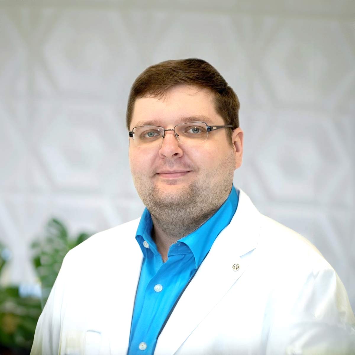 Emīls Talačka
ultrasonography specialist, radiologist
Competent and certified radiology specialist with in-depth knowledge in ultrasonography diagnostics, who helps in timely diagnosis of various diseases of internal organs and soft tissues. The doctor performs ultrasonography of the abdominal cavity, thyroid gland, kidneys, bladder and other structures, providing an accurate interpretation of the examination results that is understandable to the patient.
Emīls Talačka
ultrasonography specialist, radiologist
Competent and certified radiology specialist with in-depth knowledge in ultrasonography diagnostics, who helps in timely diagnosis of various diseases of internal organs and soft tissues. The doctor performs ultrasonography of the abdominal cavity, thyroid gland, kidneys, bladder and other structures, providing an accurate interpretation of the examination results that is understandable to the patient.
Types of ultrasound examinations at the FORTEMED medical centre:
- Abdominal cavity (liver, pancreas, gallbladder, bile ducts, spleen, etc.);
- Kidneys and urinary tract;
- Cervical lymph nodes and soft tissues;
- Thyroid gland;
- Prostate.
*General ultrasonography is performed on patients aged 14 and older. Younger patients must make an appointment for paediatric ultrasonography.
Ultrasonographic examination of joints
Ultrasonography of the musculoskeletal system aims to provide timely and accurate diagnosis of musculoskeletal diseases. Ultrasonography enables assessment of the condition of muscles, tendons, ligaments, joints, and soft tissues, and the identification of inflammatory, degenerative, or traumatic changes at an early stage. It is a safe, painless, and repeatable examination method that enables physicians to make a diagnosis and monitor the effectiveness of treatment over time.
FORTEMED experts in ultrasound examination of the musculoskeletal system

During the examination, the doctor can diagnose:
- Smooth or uneven joint contours;
- The presence or absence of loose bodies in the joint;
- Effusion or fluid in the joint;
- Inflammation of the inner layer of the joint or synovial membrane;
- Assess the condition of ligaments and tendons - their stretching, tears, inflammation, chronic changes;
- Partially assess the condition of cartilage and menisci;
- Detect cysts near the joints, such as a Baker's cyst in the knee joint.
This type of diagnosis enables accurate detection of pathologies. The advantage of ultrasound is that, if necessary, you can compare symmetrical joints.
Indications for ultrasound of the joints:
- Injuries to joints, muscles, ligaments and tendons;
- Swelling of the joints;
- Pain in the joints or surrounding tissues;
- Difficulty moving the joints;
- Suspected arthritis, bursitis, tendinitis.
Types of ultrasonographic examinations of the joints
The FORTEMED medical centre performs the following types of joint ultrasound:
- Shoulder joint ultrasound;
- Elbow joint ultrasound;
- Wrist and finger joint ultrasound;
- Knee joint ultrasound;
- Shin and foot ultrasound.
Diagnostic examination is not recommended if the patient has received an injection directly into the joint within 24 hours of the examination.
Gynaecological ultrasound
A gynaecological ultrasound is a visual examination of the pelvic organs and female reproductive system using ultrasound waves. Ultrasound is a painless and safe diagnostic method that is widely used to diagnose and detect gynaecological diseases, as well as to monitor pregnancy.
FORTEMED team of experts in gynaecological ultrasound examination


A gynaecological ultrasound is an examination that is usually an integral part of a visit to the gynaecologist. Most often, ultrasound is performed transvaginally – by inserting a probe into the vagina – but it may also be performed transabdominally – through the front wall of the abdomen.
This examination allows the doctor to assess the condition of the uterine cavity, uterine wall, uterine lining (endometrium), fallopian tubes, ovaries, bladder, cervix, and vagina, diagnose congenital or acquired pathologies and changes, and localise and measure benign or malignant tumours.
Transvaginal ultrasound should be performed in cases of unexplained vaginal bleeding, pelvic pain, infertility, suspected cysts and other changes, and to check the correct insertion of an intrauterine device.
Gynaecological ultrasound can detect diseases such as:
- Uterine fibroids;
- Cysts;
- Inflammatory diseases of the pelvic organs;
- Ectopic pregnancy;
- Cancer of the reproductive organs.
At the FORTEMED medical centre, experienced gynaecologists manage patients' health through gynaecological care and ultrasound diagnostics. The FORTEMED team of doctors offers gynaecological ultrasound examinations using the new-generation Mindrey Resona I9 high-resolution device, enabling a wide range of expert-level examinations.
After the examination and review of the results, the doctor can make a diagnosis and prescribe further steps and a treatment plan.
Important! Successful transabdominal ultrasound diagnosis requires a full bladder, i.e. it is recommended to drink 500-700 ml of fluid 2-3 hours before the examination. This examination is recommended annually, even in the absence of complaints, for preventive purposes.
Breast ultrasonography
Breast ultrasonography is a safe, painless examination that provides detailed information on the structure of the mammary glands. It is an integral part of preventive treatment, helping detect tissue changes in a timely manner and prevent the development of malignant formations. Regular ultrasound examinations help maintain breast health and give you confidence in your well-being.
FORTEMED team of experts in breast ultrasound examinations


Ultrasound helps the doctor visually assess changes in breast structure, identify cystic or tumour formations, mastitis, and other conditions that require treatment or observation.
At the FORTEMED medical centre, breast ultrasound is performed using the most modern ultrasound equipment.
The FORTEMED Medical Centre employs highly experienced breast surgeons, including Dr. Pēteris Loža and Dr. Jeļena Maksimenko, who use modern equipment to diagnose and treat all breast diseases, including benign and malignant formations.
Ultrasound diagnostics for children
Paediatric ultrasound examinations are vital, safe and painless, allowing us to assess the health of our young patients with maximum accuracy and gentleness. A major advantage of this method is the absence of radiation exposure, making it an ideal choice for younger children.
These examinations enable a detailed assessment of the condition of various organ systems and structures, including the gastrointestinal tract, kidneys, soft tissues, heart and blood vessels, lungs, and more. For effective diagnosis, inflammation, tumours, cysts, structural organ changes, and other conditions are often detected. In addition, ultrasound can assess organ function and blood flow to ensure normal function.
At the FORTEMED Medical Centre, we are proud of our ultrasound station, which is specially equipped to perform ultrasound examinations on children. It includes smaller, more delicate probes ideal for young patients' bodies, as well as specialised software that ensures high image accuracy and detail.
With a special focus on our young patients, we ensure ultrasound examinations are conducted in a friendly, relaxed environment. The highly qualified radiologist at the Children's Clinical University Hospital, Dr. Nataļja Merkla, who performs ultrasound examinations, has special training and experience working with children, which allows parents to receive comprehensive information and support. Each examination is explained in detail to parents, and the results are considered when developing the most appropriate treatment plan.